Anatomy Diagram Rib Area - Location Of The Liver Hepatitis C Trust
Anatomy Diagram Rib Area - Location Of The Liver Hepatitis C Trust. Check out the premium anatomy course to see the full version of this video and all other anatomy videos. It has a roughened area on its upper surface, from which the serratus anterior muscle originates. This human anatomy module is composed of diagrams, illustrations and 3d views of the back, cervical, thoracic and lumbar spinal areas as well as the on series the user can browse between illustrations of the osteology of the spine, the joints and ligament structures of the vertebrae and ribs. Butterflys in tummy skeleton rib cage anatomical wall hanging, art print antique vintage dictionary book page unique home decor artwork. Area between the head and the tubercle of the rib.
ads/bitcoin1.txt
The diaphragm forms the upper surface of the abdomen. Learn vocabulary, terms and more with flashcards, games and other study tools. They also have a role in. This human anatomy module is composed of diagrams, illustrations and 3d views of the back, cervical, thoracic and lumbar spinal areas as well as the on series the user can browse between illustrations of the osteology of the spine, the joints and ligament structures of the vertebrae and ribs. The ribs are elastic arches of bone, which form a large part of the thoracic skeleton.
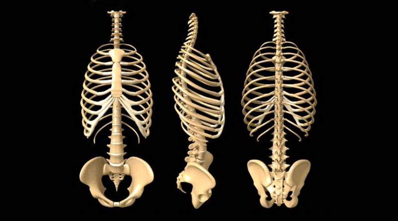
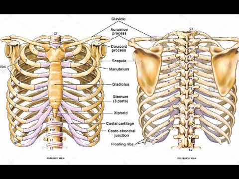
Anatomy Of The Human Rib Cage Badass Chiropractic And Wellness from drkarenhudes.ca The rib cage, shaped in a mild cone shape and more flexible than most bone sets, is made up of varying elements such as the thoracic vertebra, 12 equally paired ribs, costal cartilage, and held together anteriorly by the sternum. Interactive tutorials about the ribs and sternum bones, with labeled images and diagrams featuring the beautiful illustrations of getbodysmart. The ribs are a set of twelve paired bones which form the protective 'cage' of the thorax. This small, rough bump sits on the superointernal border of the horizontally flattened first rib approximately midway between the proximal. The diaphragm forms the upper surface of the abdomen. The human rib cage is made up of 12 pairs of ribs, some of which attach to a bony process in the front of the chest called the sternum. When examining individual rib bones, you'll notice that some have different structures, so anatomists categorize ribs rib 2 is also quite curved, but it is longer than rib one and not as flat. Area between the head and the tubercle of the rib.
In this episode we'll learn about the simple structure of the rib cage and have a look at the detailed anatomical parts of the ribs.
ads/bitcoin2.txt
Related posts of anatomy of ribs and its related area diagram of human nose diagram. Human skin cross section anatomy diagram. They articulate with the vertebral column posteriorly, and terminate anteriorly as cartilage (known as costal cartilage). 20.10.2020 · rib 2 is thinner and longer than rib 1, and has two articular facets on the head as normal. The rib cage, shaped in a mild cone shape and more flexible than most bone sets, is made up of varying elements such as the thoracic vertebra, 12 equally paired ribs, costal cartilage, and held together anteriorly by the sternum. Ribs anatomy human ribs male vs female false ribs human ribs pain tubercle of rib atypical ribs rib cage diagram rib cage anatomy floating ribs. The human rib cage is made up of 12 pairs of ribs, some of which attach to a bony process in the front of the chest called the sternum. Area between the head and the tubercle of the rib. Lessons on the bone markings of the ribs and sternum. The diaphragm forms the upper surface of the abdomen. Ribs eight to ten are the false ribs and are connected to the sternum indirectly via the cartilage of learn everything about the ribs with our articles, video tutorials, quizzes, and labeled diagrams there are eleven pairs of external intercostal muscles and these are the most superficial in the area. Rib anatomy, thoracic rib, rib bone. The serratus anterior muscle originates from a roughened area near the middle of.
They are twelve in number on either side; They articulate with the vertebral column posteriorly, and terminate anteriorly as cartilage (known as costal cartilage). They also have a role in. This entry was posted in anatomy by admin. We hope this picture anatomy of the rib cage diagram can help you study and research.
Two Minutes Of Anatomy Ribcage Youtube from i.ytimg.com But this number may be increased by the development of a cervical or lumbar rib, or may be diminished to eleven. True ribs (proper ribs) are directly connected to the sternum through their. The diaphragm forms the upper surface of the abdomen. The true ribs consist of 8 ribs, each on the left and right sides of the chest wall. Human skin cross section anatomy diagram. Interactive tutorials about the ribs and sternum bones, with labeled images and diagrams featuring the beautiful illustrations of getbodysmart. Learn vocabulary, terms and more with flashcards, games and other study tools. The long curved bones which form the rib cage.
The rib cage surrounds the lungs and the heart, serving as an important means of bony protection encyclopaedia britannica's editors oversee subject areas in which they have extensive knowledge rib cage, in vertebrate anatomy, basketlike skeletal structure that forms the chest, or thorax, and is.
ads/bitcoin2.txt
The serratus anterior muscle originates from a roughened area near the middle of. The long curved bones which form the rib cage. Butterflys in tummy skeleton rib cage anatomical wall hanging, art print antique vintage dictionary book page unique home decor artwork. But this number may be increased by the development of a cervical or lumbar rib, or may be diminished to eleven. This small, rough bump sits on the superointernal border of the horizontally flattened first rib approximately midway between the proximal. The rib cage, shaped in a mild cone shape and more flexible than most bone sets, is made up of varying elements such as the thoracic vertebra, 12 equally paired ribs, costal cartilage, and held together anteriorly by the sternum. The human rib cage is made up of 12 pairs of ribs, some of which attach to a bony process in the front of the chest called the sternum. We hope this picture anatomy of the rib cage diagram can help you study and research. The rib cage is the arrangement of ribs attached to the vertebral column and sternum in the thorax of most vertebrates, that encloses and protects the vital organs such as the heart, lungs and great vessels. The ribs are elastic arches of bone, which form a large part of the thoracic skeleton. As part of the bony thorax, the ribs protect the internal thoracic organs. Generally, ribs 1 to 7 are connected to the sternum by their costal cartilages and are called true ribs, whereas ribs 8 to 12 are termed false ribs. Types of human body joints.
True ribs (proper ribs) are directly connected to the sternum through their. It has a roughened area on its upper surface, from which the serratus anterior muscle originates. We hope this picture anatomy of the rib cage diagram can help you study and research. Each are symmetrically paired on a right and left side. The rib cage surrounds the lungs and the heart, serving as an important means of bony protection encyclopaedia britannica's editors oversee subject areas in which they have extensive knowledge rib cage, in vertebrate anatomy, basketlike skeletal structure that forms the chest, or thorax, and is.
Ribs Anatomy Types Ossification Clinical Significance How To Relief Human Body Anatomy Rib Cage Anatomy Body Anatomy from i.pinimg.com Lessons on the bone markings of the ribs and sternum. Start studying anatomy of the rib. Thoracic cage unlabeled rib cage unlabeled l. Rib bone anatomy and landmarks. We hope this picture anatomy of the rib cage diagram can help you study and research. Each are symmetrically paired on a right and left side. Interactive tutorials about the ribs and sternum bones, with labeled images and diagrams featuring the beautiful illustrations of getbodysmart. Medical human chest skeletal bone structure model.
Incredible india human body anatomy.
ads/bitcoin2.txt
When examining individual rib bones, you'll notice that some have different structures, so anatomists categorize ribs rib 2 is also quite curved, but it is longer than rib one and not as flat. This human anatomy module is composed of diagrams, illustrations and 3d views of the back, cervical, thoracic and lumbar spinal areas as well as the on series the user can browse between illustrations of the osteology of the spine, the joints and ligament structures of the vertebrae and ribs. It has a roughened area on its upper surface, from which the serratus anterior muscle originates. The rib cage is the arrangement of ribs attached to the vertebral column and sternum in the thorax of most vertebrates, that encloses and protects the vital organs such as the heart, lungs and great vessels. The human rib cage is made up of 12 pairs of ribs, some of which attach to a bony process in the front of the chest called the sternum. Each are symmetrically paired on a right and left side. The long curved bones which form the rib cage. This entry was posted in anatomy by admin. The ribs are a set of twelve paired bones which form the protective 'cage' of the thorax. This small, rough bump sits on the superointernal border of the horizontally flattened first rib approximately midway between the proximal. The rib cage is an origin and insertion area for many muscles. Types of human body joints. The first seven ribs attach directly to the.
ads/bitcoin3.txt
ads/bitcoin4.txt
ads/bitcoin5.txt
0 Response to "Anatomy Diagram Rib Area - Location Of The Liver Hepatitis C Trust"


0 Response to "Anatomy Diagram Rib Area - Location Of The Liver Hepatitis C Trust"
Post a Comment